Macrophages Become Myofibroblasts in Endometriosis Fibrosis
Dec 5, 2025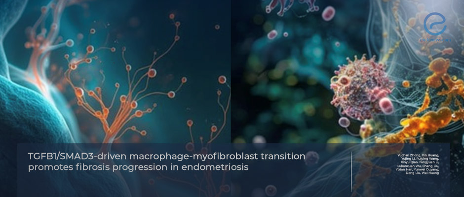
Human and mouse data uncover TGFB1/SMAD3 as a central regulator of fibrosis through macrophage reprogramming.
Key Points
Highlights:
- Endometriotic lesions exhibit fibrosis driven by TGFB1/SMAD3 activation.
- Macrophage–myofibroblast transition (MMT) emerges as a novel contributor to endometriosis-associated fibrosis.
Importance:
- Fibrosis is a defining but under-explained pathology of endometriosis.This study identifies MMT as a mechanistic link between inflammation and fibrotic remodeling.
- TGFB1/SMAD3 emerges as a central upstream regulator of fibrosis, validating it as a highly relevant therapeutic target.
What's Done Here:
- Human eutopic and ectopic endometrial samples from women with moderate–severe endometriosis were analyzed for MMT markers.
- A BALB/c mouse model of endometriosis was created and treated with inhibitors of SMAD3 phosphorylation (SIS3) or TGFB1 receptor activity (SB431542).
- Histology, immunohistochemistry, and molecular assays evaluated fibrosis, MMT, and lesion survival.
Key Results:
- Endometriotic tissues showed elevated TGFB1, SMAD3 activation, and accumulation of MMT cells defined by CD68/α-SMA positivity.
- Mouse lesions demonstrated collagen deposition and increased fibrosis.
- Blocking TGFB1/SMAD3 signaling reduced MMT, decreased extracellular matrix deposition, and impaired ectopic lesion survival.
- TGFB1/SMAD3 pathway identified as necessary for fibrotic progression and lesion maintenance.
Strength and Limitations:
- Combining human tissue, mouse models, and mechanistic interventions with multimodal design; identification of a previously unrecognized cell transition (MMT) in endometriosis fibrosis as a novelty, and the demonstration of a causal pathway involvement via targeted inhibition are the strengths of the study.
- Limited human sample size and its restriction to ovarian endometriomas, absence of flow cytometry for MMT cell quantification, EMT that was not conclusively shown in the mouse model and the mechanistic resolution of TGFB1/SMAD3-MMT requires lineage tracing and knockout models are the limitations.
From the Editor-in-Chief – EndoNews
"Fibrosis is one of the most consequential yet least understood dimensions of endometriosis biology. It shapes lesion architecture, may dictate pain severity, compromises ovarian reserve, and disrupts endometrial receptivity. Despite its overwhelming clinical relevance, the cellular origin of fibrosis in endometriosis has long remained speculative—fragmented across concepts of fibroblast activation, EMT, and chronic inflammation. This study brings rare clarity to that landscape.
The work is scientifically important because it reveals a missing cellular link: macrophage–myofibroblast transition (MMT) as an active driver of fibrosis in both ectopic lesions and the eutopic endometrium. Identifying MMT in endometriosis reshapes our understanding of lesion biology. It positions macrophages not only as inflammatory participants but as direct fibrogenic effectors capable of producing collagen and remodeling the extracellular matrix.
Equally compelling is the demonstration that this process is under the control of the TGFB1/SMAD3 axis, a pathway consistently elevated in endometriosis yet rarely connected mechanistically to the fibrotic phenotype observed in clinical practice. By showing that targeted inhibition of TGFB1 signaling suppresses MMT, reduces collagen accumulation, and even impairs lesion survival, the authors provide rare experimental evidence that fibrosis is not a passive scar—it is a regulated, targetable component of disease persistence.
The strength of this study lies in its multimodal approach—integrating human tissues, mouse models, histology, and pathway inhibition—to construct a coherent mechanistic story. Few studies in the endometriosis field achieve this degree of consistency across platforms. The findings feel scientifically “tight,” internally validated, and biologically plausible.
The implications are profound. For decades, fibrosis has been accepted as an inevitable endpoint of lesion progression, something clinicians manage surgically rather than biologically. This study challenges that assumption. If macrophage reprogramming via TGFB1/SMAD3 is a core engine of fibrotic remodeling, then interrupting this axis could alter the trajectory of disease, not merely treat its consequences. It opens the door to therapies aimed at reducing stiffness, improving decidualization, restoring receptivity, and perhaps lowering recurrence risk.
Endometriosis research has long needed mechanistic breakthroughs that unify its inflammatory, fibrotic, and survival pathways. Zhong et al. deliver exactly that: a compelling mechanistic framework—MMT driven by TGFB1/SMAD3—that connects cellular behavior to clinical phenotype. This is not just an incremental advance; it is a meaningful shift in how we conceptualize fibrogenesis in endometriosis.
Their work should stimulate a new era of investigation focused on lineage tracing, macrophage-specific modulation, and translational targeting of fibrosis. For patients, the long-term hope is clear: treatments that address why fibrosis forms, not just how to remove it once it does."
Lay Summary
Fibrosis is a major but often underrecognized feature of endometriosis. It contributes to pain, adhesions, impaired endometrial receptivity, and progressive lesion stiffness. Although fibrosis is well documented in both ectopic lesions and the eutopic endometrium, the exact cellular mechanisms driving this process have remained unclear.
In this study published in Molecular Human Reproduction, researchers led by Dr. Huan investigated whether a specific cellular transformation—known as macrophage–myofibroblast transition (MMT)—contributes to fibrosis in endometriosis. MMT occurs when macrophages begin expressing myofibroblast markers such as α-SMA and start producing collagen, effectively becoming fibrogenic cells. This phenomenon has been described in other fibrotic diseases but had not been clearly demonstrated in endometriosis.
Using human tissue samples and a mouse model of endometriosis, the researchers found that lesions with higher fibrosis also showed increased activation of the TGFB1/SMAD3 signaling pathway, a central regulator of tissue remodeling. These fibrotic lesions contained cells co-expressing macrophage and myofibroblast markers, indicating active MMT. In the mouse model, endometriotic implants showed collagen accumulation and increased expression of fibrosis-related proteins, confirming the presence of similar pathological changes.
Importantly, when the TGFB1/SMAD3 pathway was inhibited—either by blocking SMAD3 phosphorylation (SIS3) or TGFB1 receptor activity (SB431542)—both MMT and fibrotic remodeling were significantly reduced. Lesion survival also declined under these treatments, suggesting that fibrosis is not merely a byproduct of inflammation but a process that actively supports lesion persistence.
Overall, this work identifies MMT as a novel and previously underappreciated driver of fibrosis in endometriosis and positions the TGFB1/SMAD3 axis as a central regulator of this transition. These findings highlight a potentially actionable therapeutic pathway that could, in the future, help reduce fibrosis, alleviate symptoms, and limit lesion progression in patients with endometriosis.
Research Source: https://pubmed.ncbi.nlm.nih.gov/41056423/
macrophage myofibroblast fibrosis microenvironment TGF beta signal transduction collagen

