With so many imaging tools and modalities, which one should be used for endometriosis?
Sep 13, 2017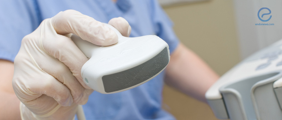
Pre-Operative Imaging tools for the Treatment of Deeply Infiltrating Endometriosis
Key Points
Highlights:
- Transvaginal ultrasonography with bowel preparation has been seen to offer improved lesion identification while rectal endoscopic sonography may be as accurate as MRI for the detection of submucosal/musical involvement of DIE.
Key Results:
- Transvaginal ultrasonography with bowel preparation has been seen to offer increased specificity and sensitivity in the visualization of rectosigmoid-involving endometriosis.
- For the visualization of endometrial involvement (depth), MRI and RES were able to predict the depth of the lesion(s) 50 and 58% of the time, respectively.
- TVUS-BP cannot evaluate anterior compartment structures such as the uterosacral ligaments, vaginal wall, rectovaginal septum.
- Radiologists trained to use TVUS-BP obtained proficiency after 70 scans.
Limitations:
- The studies included in this summary article have a small sample size. Additionally, only one radiologist was followed in the study by Young et al.
Lay Summary
Intestinal endometriosis affects 5 to 12% of women with endometriosis and commonly presents as dyschezia, rectal bleeding, cyclic defecation pain, constipation, and diarrhea. Currently, surgical treatment includes segmental rectal resection with colorectal anastomosis, but it is often riddled with complications. Thus, identifying such lesions as early as possible may lessen the severity of post-treatment complications and disease symptoms. Transvaginal ultrasonography (TVUS), rectal endoscopic sonography (RES) and magnetic resonance imaging (MRI) can be used to detect and localize intestinal endometriosis. Transvaginal ultrasonography is often used as a first-line imaging modality to identify lesions and has been seen to improve with bowel preparation. Additionally, a comparison of MRI and RES as a predictor of colorectal-involving endometriosis invasion is explained.
This study included 40 consecutive patients with histologically-proven deep infiltrating endometriosis (DIE) who underwent laparoscopic colorectal resection after pre-operative RES imaging. Out of the 40 patients with DIE include in the study, the most common symptoms reported were dysmenorrhea (80%), dyschezia (70%), chronic pelvic pain (70%), and dyspareunia (68%).
Histopathological examination revealed that 18/40 patients (45%) had muscular invasion only, 19/40 patients (48%) had submucosal or mucosal infiltration, and the remaining 3/40 patients had only serosal involvement (no deep invasion). MRI sensitivity for the detection of muscular layer invasion was 68% and specificity was 81%. In total, MRI was able to correctly predict the infiltrative depth of endometriosis 50% of the time.
RES endoscopic sonography diagnosed 14 patients with muscular invasion, 23 patients with muscular and submucosal involvement, and 3 patients with involvement of all three layers. When compared to the gold standard (histologic evaluation), 8/14 (57%) muscular invasion, 15/26 (58%) submucosal or mucosal involvement cases were correctly identified by RES. The sensitivity of RES for the detection of submucosal or mucosal invasion was 79% and specificity was 48%. In total, RES was able to correctly predict the infiltrative depth of endometriosis 58% of the time.
TVUS can be enhanced by first performing bowel preparation (TVUS-BP) to achieve similar specificity and sensitivity of MRI in detecting DIE. Ros et al. compared the accuracy of TVUS and TVUS-BP in the diagnosis of rectosigmoid DIE. 40 women underwent TVUS followed by TVUS-BP before exploratory laparoscopy to see the presence of rectosigmoid lesions. The sensitivity, specificity, and positive predictive value of TVUS with and withowereBP was seen to be 100 vs 73%, 96 vs 88%, and 94 vs 79%. While TVUS-BP improved the identification of rectosigmoid lesions, it should be noted that TVUS-BP cannot evaluate anterior compartment structures such as the uterosacral ligaments, vaginal wall, rectovaginal septum, appendix, terminal ileum, and upper sigmoid colon.
As with all other imaging tools, there is a learning curve expected of radiologists who learn TVUS-BP. A study by Young et al. measured the learning curve of a board-certified radiologist. After undergoing five days of initial observation at a high-volume clinic, the radiologist went on to perform 117 independent scans, each followed by confirmatory operative laparoscopy. During course of the study’s 37 months, the authors observed that the radiologist obtained proficiency after 70 scans.
Research Source: https://www.ncbi.nlm.nih.gov/pubmed/28866097
imaging ultrasound recto-sigmoid MRI DIE endometriosis

