"Speckle sign": a novel transvaginal ultrasound finding for posterior compartment endometriosis
Feb 4, 2021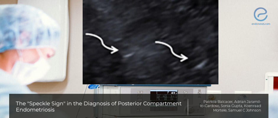
Transvaginal ultrasound examination may yield diagnostic "speckle sign" for endometriosis
Key Points
Importance:
- Improving the accuracy of posterior compartment deep infiltrating endometriosis diagnosis is important.
Highlights:
- Internal hyperechoic echoes (speckles) within the hypoechoic tissue in the posterior compartment, "speckle sign" is useful for the diagnosis of posterior compartment endometriosis, and is often found associated with other imaging features of deep endometriosis.
- "Speckle sign" may improve the diagnosis of deep infiltration endometriosis in patients with low clinical suspicion, with a high interobserver agreement.
Background:
- Deep infiltrating endometriosis extends below the peritoneum, involves the uterosacral ligaments, rectosigmoid colon, vagina, and bladder.
- Physical examination has limited value in evaluating the disease extent.
- Pelvic ultrasound is generally utilized for the assessment of ovarian endometriomas, besides revealing the fluid status of the Douglas pouch.
- Despite the presence of guidelines by uterine motility and adhesion evaluation, still, deep infiltrating endometriosis is a challenging diagnosis by ultrasound.
What's done here?
- Transvaginal ultrasound examination of 25 women with surgically and histopathologically confirmed Douglas pouch endometriosis was reassessed by two expert radiologists, unaware of clinical information.
-
Apart from the presence, size, heterogeneity, and margins of the “Speckle sign” the following
additional features of endometriosis were evaluated: -
- Adnexal endometrioma, adherent “kissing ovaries”, posterior compartment endometriosis, bowel tethering of bowel, adenomyosis, retroflex uterus, and free pelvic fluid.
- The diagnostic yield of ultrasonographic findings and interobserver agreement were calculated.
Key Results:
- The proposed “speckle sign” is often found with other ultrasound features of endometriosis is valuable in identifying posterior compartment endometriosis.
- There was a 96% agreement between two expert radiologists in identifying posterior compartment endometriosis showing this sign.
Limitations:
- There exists an inherent bias due to the retrospective nature of this study and the small sampling size of the patients without any control group existing.
- This could result in a false-positive rate of the so-called “speckled sign”. Besides, color Doppler was not included in this patient group which might be regarded as a drawback by some experts on the subject.
Lay Summary
Dr. Balcacer and associates from three medical institutions located in the USA report the results of their retrospective study on deep infiltrating endometriosis utilizing transvaginal ultrasound examination and the proposed “speckle sign” in the "Journal of Ultrasound Medicine".
Pelvic ultrasound is routinely used mainly for the diagnostic workup of endometriosis but deep infiltrating endometriosis may cause diagnostic challenges due to common atypical presentations. This study focused on defining a meaningful sign in patients with deep invasive endometriosis where the authors name as “speckle sign” depending on hyperechoic foci found in hypoechoic lesions in the Douglas pouch.
Patients with surgically and histopathologically confirmed posterior compartment endometriosis who also had a transvaginal ultrasound within 12 months of surgery were re-assessed in this study. The transvaginal ultrasound exams of 25 patients fitting these prerequisites were studied by two expert radiologists independently. There was a 96% agreement among the two expert radiologists in terms of the presence of the “Speckle sign".
“Speckle sign” searched by these experts to diagnose posterior compartment endometriosis is defined as internal hyperechoic echoes or “speckles” within the hypoechoic tissue in this anatomic region. The size, echogenicity, heterogeneity surrounding the bright echoes, and margin regularity were also tabulated along with this sign.
Authors found that the "speckle sign" is useful for the diagnosis of posterior compartment endometriosis, is often associated with other imaging features of deep endometriosis, has a high inter-observer agreement, and this sign may be helpful for improving the diagnosis of deep infiltrative endometriosis in patients with low clinical suspicion.
Color Doppler was not included in this study group. Besides the inherent bias due to the retrospective nature and small sampling size of patients without any control group yields an important necessity for future research. Future research should also include differential diagnosis of other pathologies that affect the Douglas pouch, like clots or pelvic inflammatory disease.
Research Source: https://pubmed.ncbi.nlm.nih.gov/33417291/
ultrasonography endometriosis Douglas pouch speckle sign hyper-echoic hypo-echoic endometrioma kissing ovaries

