Blood Based Multi-Omics Endometriosis Detection
May 21, 2026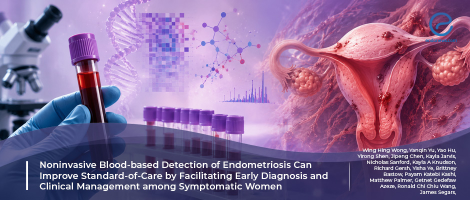
Machine learning blood test complements ultrasound and MRI in endometriosis detection
Key Points
Highlights:
- A machine learning–integrated multi-omic blood assay demonstrated high diagnostic accuracy for endometriosis across different menstrual cycle phases.
- The blood-based assay identified 61.5% of histologically confirmed endometriosis cases that were missed by ultrasound and/or MRI.
- The diagnostic model combined serum microRNAs, protein biomarkers, progesterone, age, and BMI using a random forest algorithm.
Importance:
- This study supports the emerging role of minimally invasive, blood-based multi-omic approaches as complementary diagnostic tools in endometriosis evaluation.
What’s done here?
- In this multicenter case-control study, 298 reproductive-age symptomatic women undergoing evaluation for suspected endometriosis were enrolled.
- A training cohort of 218 participants was used to develop the diagnostic model, while an independent validation cohort of 80 participants was used to assess generalizability.
- Peripheral blood samples obtained before surgery were analyzed for 3 serum microRNAs (miR-17-5p, miR-15b-5p, miR-92a-3p), 3 protein biomarkers (CA125, CA19-9, SHBG), progesterone, age, and BMI.The molecular and clinical variables were integrated using a random forest machine learning algorithm for binary classification of endometriosis.
Key results:
- In the independent validation cohort, the assay achieved an AUC of 0.944 with 80% sensitivity and 97.5% specificity.
- Stable diagnostic performance was observed across menstrual phases, with proliferative-phase and secretory-phase AUCs of 0.935 and 0.993, respectively.
- The blood assay correctly identified 61.5% of histologically confirmed endometriosis cases that had been classified as negative by ultrasound and/or MRI.
Strengths and Limitations:
- Strengths are the multicenter design, integration of multi-omic biomarkers with machine learning, phase-independent performance, and use of histopathological confirmation as the reference standard.
- Limitations are the modest cohort size, incomplete evaluation in early-stage disease, potential confounding by comorbidities, and lack of longitudinal validation.https://pubmed.ncbi.nlm.nih.gov/41791702/
From the Editor-in-Chief – EndoNews
"This study reflects a growing transformation in the preoperative evaluation of endometriosis: the transition from reliance primarily on imaging-visible disease toward biologically integrated disease assessment. While histopathological confirmation remains the definitive diagnostic standard, preoperative identification of endometriosis continues to be limited by variability in lesion visibility, operator-dependent imaging performance, and the difficulty of detecting subtle or superficial disease. The current work attempts to address this gap by combining molecular biomarkers with machine learning to capture systemic biological signatures associated with endometriosis.
Importantly, the value of this assay may not lie in replacing imaging or pathology, but in complementing existing clinical pathways. The finding that a substantial proportion of histologically confirmed cases missed by ultrasound or MRI were identified by the blood-based model highlights an area where current preoperative evaluation remains imperfect. This may be particularly relevant in patients with early, non-ovarian, or less structurally apparent disease, where uncertainty frequently contributes to prolonged delays before definitive confirmation.
Equally notable is the study’s emphasis on multi-omic integration rather than reliance on a single biomarker. The incorporation of circulating microRNAs, inflammatory and hormonal markers, and clinical variables acknowledges the heterogeneous and systemic nature of endometriosis biology. Nevertheless, caution remains necessary. The modest cohort size, potential confounding effects of coexisting gynecologic conditions, and limited assessment of early-stage disease indicate that larger prospective studies and broader external validation remain essential before clinical implementation can be considered.
Rather than representing a replacement for established diagnostic standards, studies such as this may signal the emergence of a more integrated preoperative assessment model in endometriosis—one that combines imaging, molecular profiling, clinical phenotyping, and ultimately pathology within a multidimensional framework."
Lay Summary
Research Source: https://pubmed.ncbi.nlm.nih.gov/41791702/
endometriosis diagnosis multiomic miRNAs proteins biomarkers

