Anatomic distribution of endometriosis
Nov 6, 2018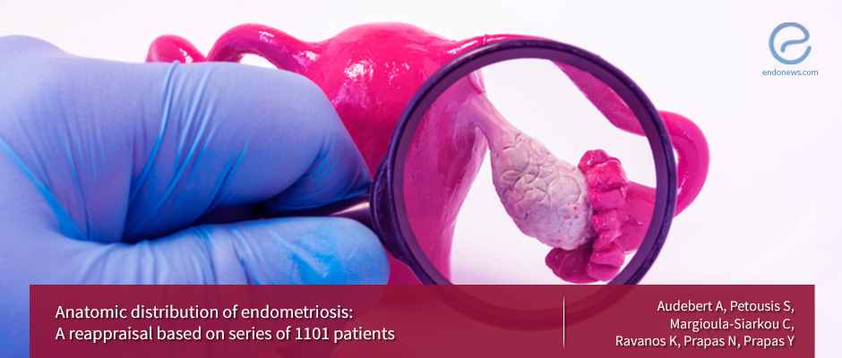
The most common site of endometriosis is the ovary.
Key Points
Highlights:
- The ovary is the most common site for endometriosis and the left side was predominant for all locations except for ovarian SI (superficial implants) and the fallopian tube.
Objectives:
- To reappraise the anatomic distribution of endometriosis lesions in cases with superficial Implants, Ovarian Endometrioma (OMA) and Deep Infiltrating Endometriosis (DIE).
What's done here:
- In this prospective observational study, 1101 women who had a laparoscopic diagnosis of endometriosis were analyzed. Subtle endometriosis lesions were searched and biopsy was performed in all cases.
- The anatomic location of the endometriosis lesions was examined as well as the rate of deep infiltrating endometriosis during the various study periods, laterality of endometriosis, pelvic adhesions’ anatomic distribution and prevalence according to past history.
Key results:
- The ovary was the most frequent site of endometriosis (in 737 patients, 66.94%); 485 presented with both OMA and SI, 227 presented with only OMA, while SI exclusively was observed only in 25 patients.
- Other frequent locations were the uterosacral ligaments (USL), the ovarian fossa, the pouch of Douglas and the bladder; the rarest being sub-mesocolic compartment (14 patients).
- DIE (excluding USL lesions except when involving the ureter) was diagnosed in 14.4% of the patients (n=159); 93.1% had associated pain, of which 89.7% had infertility as well; 77.36% had associated adhesions (n=123) and 69.9% associated OMA (n=86).
- The rate of DIE, based on laparoscopic diagnosis and histopathologic confirmation, increased progressively with a marked shift in 1995 and 2001.
- The locations involved with DIE were the rectovaginal septum (n=143), the sigmoid (n=11), the bladder (n=9) and the ureter (n=6).
- The left side was predominant for all locations except for ovarian SI and the fallopian tube. The left predominance may be due to the sigmoid colon acting as an obstacle to the diffusion of the menstrual effluent from the left fallopian tube and therefore promoting an implantation. However, the DIE lesions distribution is probably due to metaplasia of colon or Mullerian remnants metaplasia.
- Of 600 patients with pelvic adhesions, history of endometriosis was more frequent than the history of pelvic infection history of previous pelvic surgery or no history.
- The most frequent anatomic location of adhesions was adnexa (47.4%), followed by sigmoid, pouch of Douglas and uterus.
Limitations:
The limitations of this study are the bias related to factors influencing recruitment and the bias is due to some arbitrary criteria selected for lesion site coding.
Lay Summary
In the study of Audebert et al. which has been published in the journal European Journal of Obstetrics and Gynecology, the anatomic distribution of endometriosis, the rate of deep infiltrating endometriosis during the various study periods, laterality of endometriosis, pelvic adhesions’ anatomic distribution and prevalence according to past history were analyzed.
1101 patients diagnosed with endometriosis through laparoscopy procedure were included in the study. The mean age of the 1101 patients was 33.06 years (range 15-63 years). Pain, infertility or both were the most frequent complaints leading to performing the laparoscopy. A total of 3416 site-specific lesions (excluding adhesions) were noted for the 1101 patients (mean number of lesions per patient 3.10).
The most common site of endometriosis was found to be the ovary, followed by the USL, fossa ovarica, pouch of Douglas, the bladder, DIE (all sites), the uterus, the sigmoid, the fallopian tube and the round ligament respectively. The most common site of endometriosis was in correlation with the literature while the second most common site was not (the posterior broad ligament is mentioned in the literature).
DIE, of which the rate was increased progressively by the years with a marked shift in 1995 and 20001, was identified in 14.44% of the patients. It is not certain whether the incidence of DIE has progressively increased over years or whether the diagnosis facilitated than in the past by better education of reproductive physicians and the progress of technology.
Evaluation of laterality indicated a left predominance for the majority of sites: OMA (56.3%), fossa ovarica (67.50%), USL (60.71%) and bladder (57.76%). However, diaphragmatic endometriosis is exceptional, in which the study of Vercellini et al. the right side was indicated as the predominant location. The left predominance of the majority of endometriosis lesions may be due to the sigmoid colon acting as an obstacle to the diffusion of the menstrual effluent from the left fallopian tube and therefore promoting an implantation. However, the DIE lesions distribution is probably due to metaplasia of colon or Mullerian remnants metaplasia.
Adhesions were found in 54.5% of the cases and adnexa was the commonest site affected. Of patients with pelvic adhesions, history of endometriosis was more frequent than the history of pelvic infection history of previous pelvic surgery or no history.
Further prospective observational studies are needed to further elucidate on whether the increase in DIE is true as well as examining the final outcome of endometriosis after laparoscopic surgery mainly on fertility status.
Research Source: https://www.ncbi.nlm.nih.gov/pubmed/30240947
endometriosis anatomic distribution ovary deep infiltrating laterality

