The Molecular Profile Unique to Endometriosis
Jan 7, 2020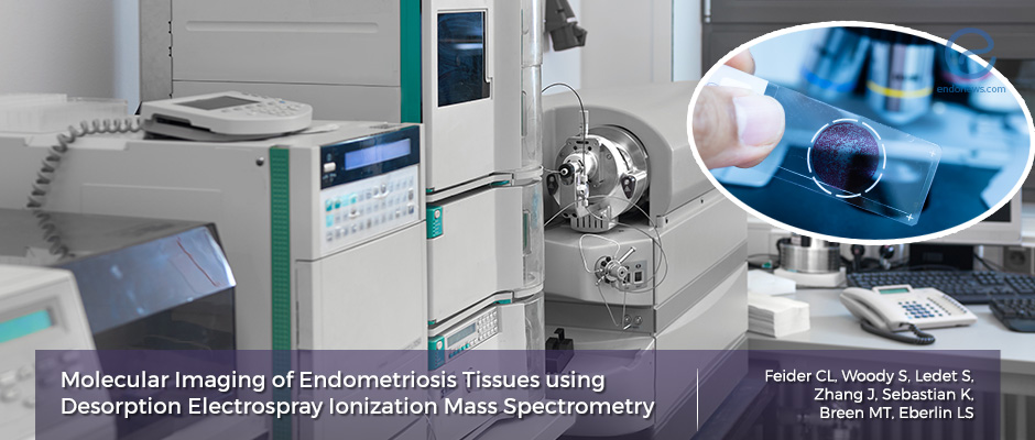
What Molecules Are Distinctively Found Among Endometriosis Tissue?
Key Points
Importance:
- This manuscript offers data on the molecular profile of endometriosis in order to gain more insights into the biomolecular alterations that may reflect the development of endometriosis.
Highlights:
- Using mass spectrometry and ionization analysis techniques, researchers have found that there are many small molecules including free fatty acids (FFAs), sphingolipids, and glycerophospholipids that are differentially seen among hundreds of collected endometriosis samples not seen in normal, eutopic endometrial tissue.
What’s done here:
- Researchers aim to investigate and discuss the need for new statistical approaches to identify different clinical, biochemical, or genetic-epigenetic abnormalities in a given endometriosis population.
Key Results:
- Molecular differences were observed both among tissue samples during inter-and intra-patient analyses, suggesting a possible biological basis for these observed differences.
- The number of distinct molecular features found after excluding isotopes and duplicates were 34, most of which being fatty acids.
- Fatty acids (especially short-chain FAs) were more characteristic of endometriosis while longer fatty acid chains with higher levels of unsaturation were more characteristic of eutopic endometrium.
- Specifically, the authors found that there was an increased amount of glycero-phosphatidic acid and glycero-phosphoglycerol species within endometriosis samples while eutopic endometrium contained more glycero-phophoinisitol species.
- Other than fatty acid metabolites, authors also found a number of small metabolites that were more characteristic of endometriosis. Hexose in its chlorine adducted form was a feature that was more indicative of endometriosis tissue.
Limitations:
- As the others suggest in the article, a true biologic process that accounts for the molecules seen in endometriosis is still unclear. This is further complicated by the fact that some patients included in this study were on hormone treatment at the time of sample collection and the fact that normal endometrium was collected from patients with proven endometriosis. As such, further study is necessary to validate the findings presented in this study.
Lay Summary
Endometriosis remains to be a disease entity that is not fully understood. However, recent studies suggest that endometriosis is characterized by several dysregulated biochemical pathways that allow for its survival and growth outside the uterine endometrium. Authors have recently published an original study that aims to analyze the molecular profile of endometriosis using mass spectrometry (MS) and ionization analysis techniques (DESI). It is through this technology that researchers aim to investigate the molecular alterations of endometriosis in the hopes of learning more about its development and findings novel biomarkers.
The study initially included a total of 269 endometriosis tissue samples that were collected at the time of laparoscopic surgery from 89 patients. No patient exclusion criteria were used in the study and the samples were collected from a variety of intrabdominal organ surfaces. 25 eutopic endometrial tissue was also collected in order to compare these results. However, 22 out of 25 eutopic and 76 out of 196 ectopic endometriosis samples from 51 patients were ultimately used for statistical analysis due to poor tissue quality. DESI-MS was used to identify small metabolites and lipids in these tissues including free fatty acids (FAs), sphingolipids such as ceramides, and glycerophospholipids. Multiple pixel data demonstrated that the endometriosis tissue contained many distinct molecular data points from those of eutopic endometrium. Likewise, endometriosis samples from several organs seem to have distinct profiles from those of eutopic endometrium.
Additionally, pathologic determination of the tissue samples to differentiate the molecular information from glandular and stromal components of the tissue samples were applied in the analysis. Important to note is that the distribution of molecular signals across women who were at varying stages of their menstrual cycle were similar in eutopic endometrium and endometriosis samples. While many molecular differences were found, the authors used another analytical method to parse out “interesting” signatures between the groups. These included small metabolites such as hexose in its chlorine form which was seen among many of the endometriosis samples. Additionally, recent studies have also suggested that based on the location of Endometriosis, specific subtypes may exist that reflect unique pathologic and molecular profiles. Thus, the authors also analyzed the molecular profile of endometriosis based on their lesion location and classified them into peritoneal, deep infiltrating endometriosis, and ovarian to see if their analysis offered any insight into possible molecules that delineate these entities. However, their analysis across all patients failed to show a significant separation or clustering based on the primary location of endometriosis.
Many aspects of this study confirm previous research that suggests a differential molecular profile of endometriosis when compared to normal endometrial tissue with the use of MS-DESI analysis. Whether the molecules found in this study will be of use in terms of biomarker development or novel drug development is uncertain. However, it is a tool that has great potential as it can break down the basic, biomolecular profile of specific cells within a tissue sample to be studied due to its high spatial resolution.
Research Source: https://www.ncbi.nlm.nih.gov/pubmed/31666535
MS MALDI-TOF pathology molecular analysis endometriosis

