PAX8 immunohistochemistry in the diagnosis of Extragenital Endometriosis
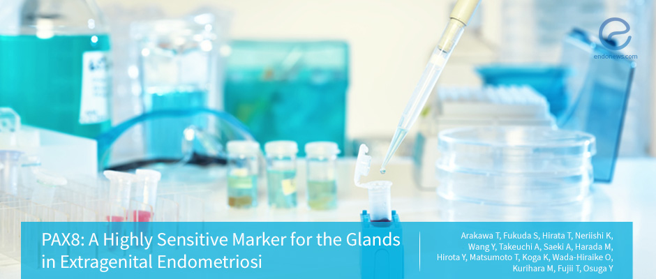
This study is performed by Arakawa T et al., from the University of Tokyo, Tokyo, Japan, and the results are published in Reproductive Sciences. Endometriosis is seen in many organs including peritoneum, ovaries, bowel, bladder, and even lung. Histopathologic diagnosis may…
Key Points Lay SummaryExtragenital endometriosis and ultrasound
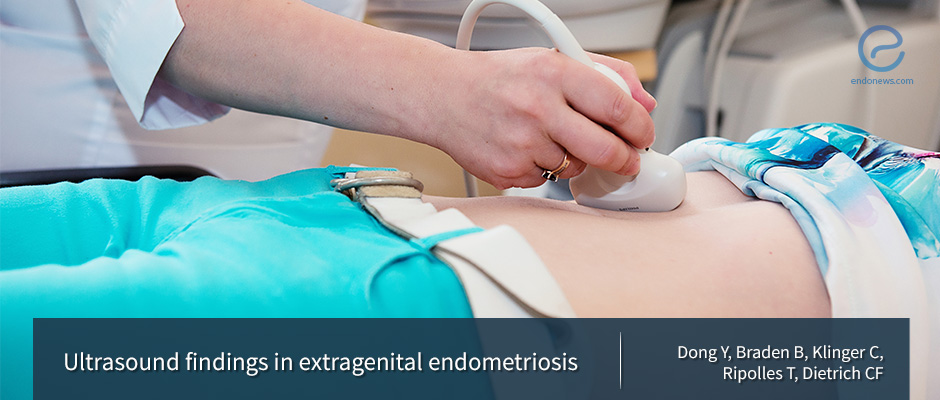
Contrast-enhanced ultrasound (CEUS) is a non-invasive imaging method with no exposure to radiation. There are a few reports on the use of CEUS in the assessment of endometriosis in the literature. Dong et al. reported 3 histopathologically proven cases of…
Key Points Lay SummaryHigh-Mobility Group Box 1 Expression Could Lead to Disease Progression
Endometriosis can often be characterized by the aberrant ectopic growth of endometriotic stromal cells (ESCs); however, research has yet to elucidate the mechanism driving this growth. The authors of this study, namely Shimizu et al., believe that the high-mobility group…
Key Points Lay Summary
 By Serdar Balci

 By Irem Onur

 By Kasthuri Nair