Imaging of gastrointestinal endometriosis
Apr 14, 2020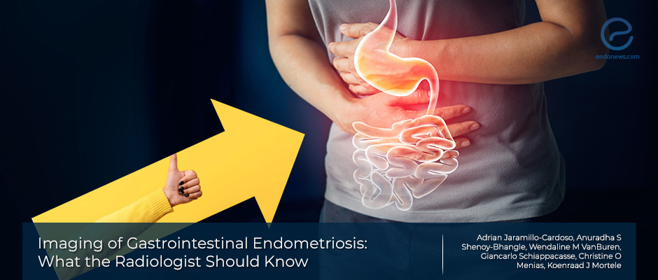
How Radiologists can best detect and characterize image findings of gastrointestinal endometriosis
Key Points
Importance:
- Often, endometriotic lesions of the gastrointestinal tract are mistaken for malignancy. It is vital to consider the presence of endometriosis in patients with relapsing abdominal pain.
Highlights:
- Ultrasound and magnetic resonance imaging are the key non-invasive imaging modalities for the initial assessment of endometriosis, although the histopathologic confirmation remains the gold standard for diagnosis.
- The key radiologic findings for ultrasound and MRI associated with endometriosis involving the stomach, small bowel, appendix, and colon should always be kept in mind.
What’s done here:
- This is a review paper aimed at highlighting the most important radiologic findings of gastrointestinal endometriosis that should be assessed by the diagnostic radiologists.
Key Results:
- Important findings to evaluate in a radiologic study for GI endometriosis include the location, number, degree of involvement, and the distance to key GI structures.
- Correct information will aid surgeons plan an effective surgical intervention for patients to benefit from surgery.
- Routine trans-abdominal US has no role in the diagnosis of gastric endometriosis. On the endoscopic US, endometriosis appears as submucosal hypoechoic nodules with irregular margins and scattered echogenic foci.
- Ultrasonography has a very limited role in detecting small bowel endometriosis.
- CT enterography has been used to demonstrate mural imaging findings associated with endometriosis such as tethering, annular thickening of the bowel, and plaque-like lesions.
- Magnetic resonance enterography has also been used to assess the small bowel wall with the advantage of not producing ionizing radiation. On MRI, these lesions are seen as a loss of the normal T2 hyperintense signal of the bowel wall, nodular and irregular thickening.
- Appendiceal endometriosis is often not detected before it causes symptoms, most commonly in the form of acute appendicitis. On the US, nodular hypoechoic lesions or irregular thickening of the wall of the appendix may be seen. On CT, a hypodense soft tissue mass or focal nodules within the appendiceal body and an enlarged appendix may be seen.
- Rectosigmoid (RS) involvement is the most common form of GI endometriosis in patients with DIE. Most commonly, transvaginal US is often used as the first-line test in the imaging of the posterior compartment in suspected cases of GI endometriosis.
- MRI has recently been suggested to offer comparable utility compared to TVUS (sensitivity and specificity >90%) for the detection of rectosigmoid endometriosis.
Limitations:
- This manuscript is a review paper that often includes authors’ experiences regarding rare forms of endometriosis (e.g. gastric endometriosis) and is thus not intended to create patient guidelines for screening or imaging.
Lay Summary
Endometriosis is a disease characterized by the ectopic implantation of endometrial tissue to surrounding pelvic and extra-pelvic structures. One frequent site of involvement is the gastrointestinal(GI) tract, which occurs especially among women with deep infiltrating endometriosis.
Most cases of GI-involving endometriosis are found around the rectosigmoid colon, up to 90%; however other sites have also been seen. While histopathologic confirmation after surgical sampling remains the gold standard, ultrasound and magnetic resonance imaging are increasingly being used for screening due to their non-invasive nature. Unfortunately, some of these endometriotic lesions are misdiagnosed as malignancy. Thus, it is critical that the Diagnostic Radiologist is informed regarding important findings that should be assessed to best aid appropriate surgical interventions if warranted.
Gastrointestinal endometriosis involving the proximal portions of the tract, including the stomach and small bowel are seldomly reported in the literature. Thus, the authors recently published this review in Abdominal Radiology to highlight what findings are important to mention in a patient’s US or MRI study. The authors then describe key image findings by specific GI segments involved, from proximal to distal, starting with the stomach, then small bowel, appendix, and rectosigmoid.
Gastric involvement is one of the least common manifestations of endometriosis but should be assessed in women with upper GI pain that is chronic or cyclic in nature. Routine trans-abdominal US has no role in the diagnosis of gastric endometriosis, however, endoscopic ultrasound has been used to report patients with this entity. Small bowel endometriosis is often difficult to detect in routine pelvic MRI studies due to the limited field of view used to obtain routine images.
For patients with a history suggestive of GI involvement, a larger field of view and coronal T1 and T2-weighted sequences should be obtained for optimal diagnosis. CT enterography has been used to demonstrate mural imaging findings associated with endometriosis such as tethering, annular thickening of the bowel, and plaque-like lesions. Magnetic resonance enterography has also been used to assess the small bowel wall with the advantage of not producing ionizing radiation. On MRI, these lesions are seen as loss of the normal T2 hyperintense signal of the bowel wall, nodular and irregular thickening which may be concurrently seen with adjacent tethering. When foci of hemorrhage are present within these lesions, they are seen as punctate, hyperintense foci on fat-suppressed pre-contrast T1W images, increasing the suspicion of the presence of endometriosis.
Appendiceal endometriosis is another uncommon entity that is often associated with patients with advanced stages of endometriosis and multicentric involvement, large right-sided endometriomas, and bladder or colon involvement. Often, symptoms do not develop (most commonly in the form of acute appendicitis) unless endometriosis has invaded the deeper, muscular layer of the appendix. Like other involved locations, AE is seen as nodular hypoechoic lesions or irregular thickening of the wall of the appendix in the US. On CT, a hypodense soft tissue mass or focal nodules within the appendiceal body and an enlarged appendix may be seen.
Rectosigmoid (RS) involvement is the most common form of GI endometriosis in patients with deeply infiltrating endometriosis. Most commonly, transvaginal US is often used as the first-line test in the imaging of the posterior compartment in suspected cases of GI endometriosis. Involvement is often seen as a nodular or plaque-like deposit that is hypoechoic compared to the muscular layer and punctate hyperechoic foci. Endometriosis involving the rectum often shows retraction and adhesions attaching the anterior rectal wall to the posterior vaginal wall or solid, focal, and tubular avascular lesions with tapering towards one end, named the comet sign.
A dynamic sign named "the sliding sign" for TVUS when no movement was seen should suggest obliteration of the pouch of Douglass due to adhesions. The transrectal US have similar findings and is sometimes performed because it gives an opportunity for direct tissue sampling via biopsy.
MRI has recently been suggested to offer comparable utility compared to TVUS (sensitivity and specificity >90%) for the detection of rectosigmoid endometriosis. These imaging modalities have the advantage of assessing lesions that may possibly involve the vesicovaginal and rectovaginal spaces which may be a limitation in laparoscopy. On MRI several signs including the fan-shaped or mushroom cap sign are seen in addition to the previously mentioned characteristics of endometriotic lesions on MRI.
Research Source: https://pubmed.ncbi.nlm.nih.gov/32236651
imaging radiology gastrointestinal endometriosis ultrasound MRI surgery

