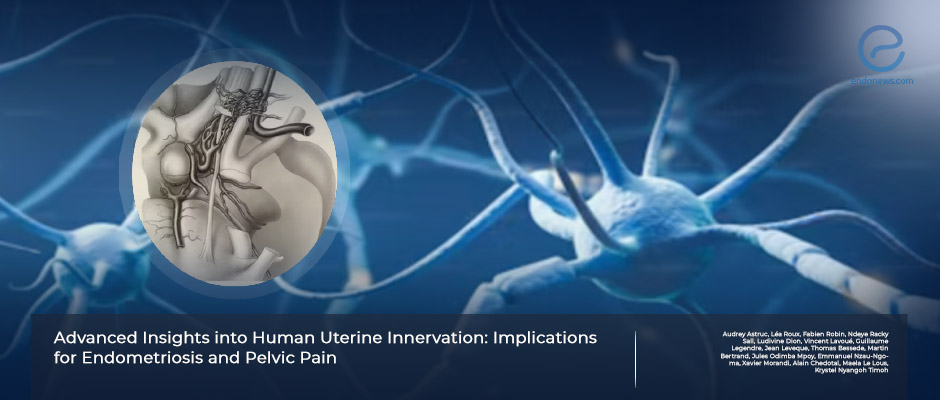
Uterine innervation and its impact on endometriosis related pelvic pain.
May 30, 2024
The cause of chronic pelvic pain in endometriosis can be resolved by examining the anatomy of uterine innervation.
Key Points
Importance:
- The anatomy of uterine innervation must be better evaluated to understand the cause of chronic pelvic pain in endometriosis.
Highlights:
- The inferior hypogastric plexus is the "commander in chief" in the pelvic and perineal regions, and any damage to it can lead to significant surgical complications.
What's done here:
- A French author team penned this review to investigate the implications of uterine innervation concerning the pathogenesis of endometriosis-related pelvic pain.
- An English literature search according to PRISMA was conducted by two authors using the keywords "uterine innervation" and " uterine anatomy."
- The complex architecture of uterine innervation providing insight into the mechanisms of pelvic pain besides revoluting surgical management are summarized.
Basic Outlines:
- The parametrium is the site of the inferior hypogastric plexus-uterine artery, and ureter, which makes the region critical to understanding various pelvic pathologies and surgical approaches.
- The inferior hypogastric plexus is typically positioned under the deep uterine vein and further extends the lateral wall of the rectum, then courses through the cervix and vagina.
- The density of inferior hypogastric plexus fibers tends to reach the highest concentration both in the vaginal fornix and paracervix.
- The sympathetic and parasympathetic nerve fibers are located in the medial side of the vesical veins.
- Uterine arteries are also highly innervated, and sympathetic nerve fibers are predominantly located in the media-adventitial border of the arterial wall to regulate the inferior hypogastric plexus vascular function.
- The middle rectal artery frequently intersects the inferior hypogastric plexus before merging into the rectal wall, which makes this feature important for surgical navigation.
- In women with endometriosis, an elevated presence of neuropeptide Y and vasoactive peptide fibers is detected within the functional layer of the endometrium. On the contrary, adenomyosis is characterized by a decrease or absence of nerve fibers.
Lay Summary
Advancements in immunochemistry, fluorochromes, computer-associated anatomical dissection, and 3D reconstruction of uterine micro-innervation brought substantial progress in pelvic neuroanatomy. The complex uterine innervation and its interactions with uterine parts and layers are yet fully revealed.
Austruc et al., from the Faculty of Medicine of Rennes University, France, reviewed the literature on the complexity of uterine innervation to deepen our understanding of female pelvic pain. The authors' final aim was to uncover valuable insights for enhancing pelvic nerve-sparing techniques in endometriosis surgery to improve surgical outcomes and quality of life.
Among 914 database searches, 45 were included in the study review. Uterine macro innervation belongs to the inferior hypogastric nerve, so the purpose was to elaborate on its input, output, location, shape, and functional roles.
The pelvic plexus, also known as the inferior hypogastric plexus, is formed by several nerves, such as the hypogastric, pelvic splanchnic, and sacral splanchnic. The description of the location of the inferior hypogastric plexus was important due to understanding surgical procedures. The strategic situation of the inferior hypogastric plexus is lateral to the rectum and vagina beneath the point where the uterine artery intersects with the ureter in the pararectal fossa. It has a bilaterally symmetric fan-shaped quadrangular formation. The junction, which the inferior hypogastric plexus usually intersects by the middle rectal artery just before it merges into the rectal wall, is a crucial anatomic landmark and essential for surgical navigation.
The inferior hypogastric plexus receives its sympathetic innervation mainly from the sacral ganglia S2 to S4 and parasympathetic innervation from the pelvic splanchnic nerve. The pain transmission pathway predominantly follows the parasympathetic route via pelvic splanchnic nerves. The dense parasympathetic fibers are located at the fallopian tube's utero tubal junction and isthmus. The uterine body shows fewer parasympathetic fibers, and nerve fiber density inside the uterus declines with age. On the other hand, sympathetic fibers of the inferior hypogastric plexus control smooth muscle sphincters and inhibit peristalsis which causes the need for a urinary catheter and challenges in spontaneous micturition if nerve-sparing techniques are not applied during hysterectomy.
In deep infiltrating endometriosis, rich innervation of sensory C, cholinergic, and adrenergic nerve fibers is found. Additionally, the high concentration of VIP neurotransmitters in the endometrium and involvement in inflammation could explain the pain associated with endometriosis.
The authors concluded that uterine innervation in endometriosis and adenomyosis presents a complex interplay of factors. Further investigations need to understand their possible roles in pain pathophysiology. This interesting review was recently published in the Journal of Clinical Medicine.
Research Source: https://pubmed.ncbi.nlm.nih.gov/38592287/
chronic pelvic pain anatomy uterine innervation hypogastric plexus pelvic plexus sacral splanchnic nerves VIP neurotransmitter endometriosis.

