Transvaginal Ultrasound and the Imaging of Endometriosis
Jul 2, 2019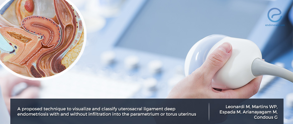
Proposed Technique for the imaging of Uterosacral ligament-involving Endometriosis using Transvaginal Ultrasound.
Key Points
Highlights:
- A proposed classification system for the "ultrasound imaging of endometriosis" may help proper evaluation for surgical decision and planning.
Importance:
- The proposed standardized classification system of endometriosis involving the uterosacral ligaments aims to help uniformity for diagnosis and may increase its diagnostic sensitivity and specificity.
What's done here:
- Authors propose a classification system that depends on the depth and location of uterosacral ligament-involving endometriosis.
Key Results:
- Type 0 denotes nodules confined to the uterosacral ligament with no evidence of infiltration to adjacent structures.
- Endometriosis involvement of >50%> of the uterosacral ligament or uterine torus, Type 1 or 2, subdivided by “T” or “P”.
Limitations:
- The proposed classification system requires further study in order to assess if there is any clinical benefit.
Lay Summary
Ultrasonography (US) is an imaging tool that uses sound waves to produce a two-dimensional picture inside of the body. The ultrasonography has become especially useful in the assessment of gynecologic pathologies such as endometriosis.
Depending on the severity of endometriosis, various intra and extra-pelvic locations may be involved. One such category of endometriosis is deep endometriosis, which is characterized by the involvement of the posterior pelvic structures such the uterosacral ligaments, structures which attach the posterior aspect of the uterus to the sacrum. Others are lateral structures such as the parametrium, and anterior structures such as the bladder. Depending on the involved location, surgical planning of endometriosis will be different.
On transvaginal US (TVUS), endometriosis is most commonly seen as a nodule. Endometriotic nodules that involve the uterosacral ligaments can be seen on TVUS but do not detect all the lesions. The sensitivity is 67% and the specificity is 86%.
Since TVUS is a very user-dependant and non-invasive diagnostic test, the authors, Leonardi et al., from Australia, propose a detailed TVUS technique guide to both standardize the way in which images are obtained and interpreted for the assessment of endometriotic nodules involving the uterosacral ligament. This uniform evaluation may help to increase sensitivity and the specificity of the endometriosis diagnosis.
In addition to the proposed technique to follow for the identification of the uterosacral ligament, the proposed classification system relies upon the location of endometriosis and the percentage of the depth of infiltration in this anatomic structure.
According to this proposed nomenclature, Type 0 denotes nodules confined to the uterosacral ligament with no evidence of infiltration to adjacent structures. Based on the involvement of the adjacent structures by >50%>, the nodule will be denoted as Type 1 or type 2, and the involvement of either uterosacral ligament or uterine torus, the subdivision into “T” or “P”.
While this nomenclature does not define all of the possible sites of endometriosis involvement, authors believe it may hold utility in the uniformity of diagnosis that will help increase in sensitivity and the specificity of the technique and will help preoperative planning to treat patients who suffer from endometriosis.
Research Source: https://www.ncbi.nlm.nih.gov/pubmed/31008537
endometriosis imaging ultrasound TVUS uterosacral ligament uterine torus

