The largest series of diaphragmatic endometriosis
Mar 23, 2022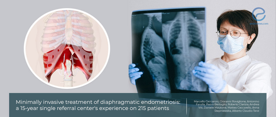
An algorithm to classify diaphragmatic endometriosis and treatment modalities with the help of 15 years’ experience
Key Points
Highlights:
- It is crucial to standardize the diagnosis, appropriate approach, and surgical technique for diaphragmatic endometriosis.
Importance:
- This study is the largest series of diaphragmatic endometriosis (DE) patients which has let to classify lesion types and make a diagram about how to treat them.
What's done here:
- The charts of 215 patients who underwent laparoscopy or video thoracoscopy, and have been diagnosed with DE in the last 15 years were evaluated.
- Diagnosis of DE has been done by the intraoperative visual finding and on histopathological examination of all specimens.
- The symptomatology, pain scores, and medical/surgical histories of patients were also taken under investigation.
- The laparoscopies were done under standardized conditions and all lesions were classified as “Foci” for bidimensional superficial lesions, “Nodules” for solid, tridimensional implants, associated with partial or full-thickness infiltration of underneath muscle layers, and “Plaques” for fibrotic bidimensional lesions, superficially infiltrating and thickening the diaphragmatic peritoneum.
- Superficial lesions were vaporized with either Argon Beam Coagulator or diathermy coagulation. The lesions larger than 1 cm were excised with peritoneal stripping. In the case of full-thickness invasion, the thoracic cavity approach was also added.
- In patients with recurrent pneumothorax, talc pleurodesis was routinely performed.
Key Results:
- Dysmenorrhea and dyspareunia as the most frequent pelvic symptoms.
- Not all the DE patients complained of diaphragmatic or thoracic symptoms, only one-fourth had thoracic symptoms.
- Only 15.4% of the DE lesions have been preoperatively seen by imaging techniques.
- More than half (58%) of patients had previous endometriosis surgeries.
- The pelvic ASRM staging revealed more than 90% as stage IV; 8.37% stage III and 1.39% stage II.
- DE lesions were mostly localized on the right side of the diaphragm (91%).
- The lesion type distribution was found to be "foci" in 62%, "plaques" in 11%, and "nodule" in 27%; more than half were treated with Argon Beam Coagulator (ABC).
- Postoperative short term follow-up showed complete pain relief and, in the long term follow-up no patients presented with recurrent DE or recurrent symptoms
Lay Summary
Diaphragmatic endometriosis is one of the most common extra pelvic endometriosis types, yet the correct incidence is unclear.
In the study conducted by Ceccaroni Marcello et al from the International School of Surgical Anatomy, Verona, Italy, the authors analyzed the data of 215 patients diagnosed with DE in the last 15 years. The lesion symptomatology of all patients, pelvic staging, DE lesion types, and type of surgeries applied were evaluated.
For typing, lesions were classified as “Foci” for bidimensional superficial lesions, “Nodules” for solid, tridimensional implants, associated with partial or full-thickness infiltration of underneath muscle layers, and “Plaques” for fibrotic bidimensional lesions, superficially infiltrating and thickening the diaphragmatic peritoneum. Results showed that the most common lesion type on the diaphragm was small foci. There was always a pelvic disease coexisting and the most common ASRM stage was the advanced stage with the most common symptoms as dysmenorrhea and dyspareunia.
Only 24.6% of patients with DE lesions had diaphragmatic or thoracic symptoms. Still, all the lesions observed were treated surgically and Argon Beam Coagulator was the most common surgical technique that was used.
All patients were put under a minimum of 3 months of postoperative hormonal treatment according to their fertility desire. The short- and long-term follow-up results were reported as total relief in all patients. The authors concluded as DE patients should be treated in specialized centers with standardized techniques to see desirable follow-up results.
Research Source: https://pubmed.ncbi.nlm.nih.gov/33398589/
thoracic endometriosis flowchart vats

