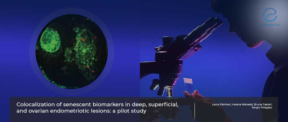
Creating a reliable indirect immunofluorescence protocol as a promising diagnostic tool for endometriosis
Jan 11, 2023
Indirect immunofluorescence staining and colocalization of the proteins p16 and lamin b1 may be useful in the diagnosis, study shows
Key Points
Highlight
- Co-localization of certain biomarkers by indirect immunofluorescence seems promising to be used as a non-invasive diagnostic tool for endometriosis.
- p16 and lamin b1 emerge as biomarkers to be used for this purpose.
Importance
- Comprehensive knowledge of cellular functions in diseases with unknown pathophysiology such as endometriosis is important in the investigation and development of new diagnostic tools.
- The similarities in the inflammatory and oxidative states of cellular senescence and endometriosis could be used as advantages in defining biomarkers.
What’s done here
- This is a qualitative descriptive study that aims to raise awareness about the colocalization of senescence-related proteins in endometriosis.
- Different endometriotic lesions such as deep infiltrative, superficial peritoneal, and ovarian were examined.
- Eutopic endometrium and lesional tissues of patients who were diagnosed with endometriosis and control tissues of patients who did not have endometriosis were included.
- Indirect immunofluorescence was performed on these tissues to target the colocalization of p16 and lamin b1 with E-cadherin proteins using a positive control, a negative control, and the tissue of interest and the staining protocol was shared in detail.
Key results
- It was previously shown that the expression of lamin b1 was decreased and p16 was increased in deep infiltrating endometriosis when compared to the eutopic endometrium.
- p16 labeling colocalized with E-cadherin in non-endometriotic, eutopic endometriotic, and deep infiltrative endometriotic tissue samples.
- E-cadherin was overexpressed in the epithelial compartment in these tissues.
- Lamin b1 had increased labeling in the same tissues even when co-localized with E-cadherin.
Limitations
- The staining patterns in the samples were interpreted visually.
- Subjective visual analysis is highly susceptible to bias.
Lay Summary
Owing to its inflammatory nature, endometriosis results in the overproduction of reactive oxygen species (ROS) which disrupt the cellular functions and eventually lead the cell to apoptosis and cell cycle arrest. Senescence, the natural state of irreversible cell cycle arrest, induces inflammation and production of ROS just like in endometriosis. The biomarkers that are used in evaluating cellular senescence are shown to be p16 and lamin B1 and the method used for this purpose is indirect immunofluorescence.
Palmieri et al. showed in a previous study that expression of lamin b1 was decreased and p16 was increased in deep infiltrating endometriosis when compared to the eutopic endometrium. The athors then performed a qualitative descriptive study aiming to raise awareness of the knowledge gap in the literature about the colocalization of senescence-related proteins in different endometriotic lesions and focusing on standardizing an immunofluorescence protocol for endometriotic lesions. The study was published in the October 2022 issue of the journal Scientific Reports.
Colocalization can be defined as the demonstration of the relationship between two or more biomarkers and the co-occurrence within the same cell or region in fluorescent microscopy. The researchers included eutopic endometrium and lesional tissues of patients who were diagnosed with endometriosis and control tissues of patients who did not have endometriosis. The endometriotic lesions belonged to patients with superficial peritoneal, deep infiltrative, and ovarian endometriosis. After collecting the tissues intraoperatively, indirect immunofluorescence was performed to target the colocalization of p16 and lamin b1 with E-cadherin proteins using a positive control, a negative control, and the tissue of interest.
The study results showed that p16 labeling colocalized with E-cadherin and E-cadherin was overexpressed in the epithelial compartment in non-endometriotic, eutopic endometriotic, and deep infiltrative endometriotic tissue samples. Moreover, lamin b1 had increased labeling in these tissues even when co-localized with E-cadherin proving the presence of glandular components.
The authors then discuss the necessity for good-quality immunofluorescence staining in detail and add that “to ensure successful immunofluorescence, critical parameters must be met” starting with fixation and permeabilization. They added that both the direct and indirect methods can be used however the indirect method has higher sensitivity. It was recommended that an experienced pathologist should review the staining patterns and evaluate co-localizations.
To conclude, the authors state that a reliable immunofluorescence protocol could be useful in defining biomarkers to be used as non-invasive diagnostic tools for endometriosis.
Research Source: https://pubmed.ncbi.nlm.nih.gov/36241900/
endometriosis indirect immunofluorescence fluorescent microscopy biomarker p16 lamin b1 senescence

