Chasing gold to find endometriosis.
Aug 12, 2022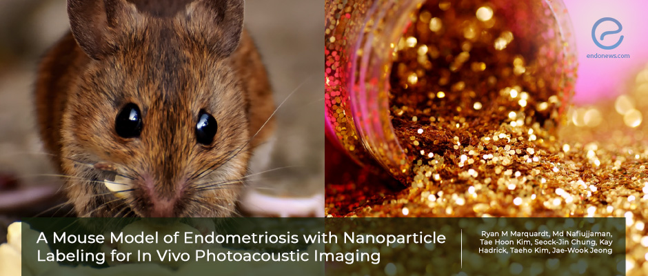
Gold nanoparticles for imaging endometriotic mouse models bring hope for future human applications.
Key Points
Highlights:
- Photoacoustic imaging of silica-coated gold nanoparticles that are bound to endometriotic lesions provides noninvasive detection of endometriosis in mice.
Importance:
- The study aimed to produce a mouse model of endometriosis, it lights a candle of hope for human applications for endometriosis diagnosis.
What's done here:
- The researchers conducted a mouse model by using silica-coated gold nanoparticles to visualize endometriotic lesion formation under photoacoustic imaging.
- They injected donor mice with estradiol for 3 days and after euthanasia of those mice, the uterine tissue have been taken out and chopped.
- The uterine tissue pieces were mixed up with fluorescent enhanced silica-coated gold nanoparticles for 3 hours.
- The prepared mixture was injected into the peritoneal cavity of recipient mice with a small incision on the abdominal wall.
- After waiting for 28 days, the endometriotic lesion formation was visualized under fluorescent microscopy and photoacoustic (ultrasound-guided) imaging.
- After sacrification of the recipient mice, the lesions were confirmed histopathlogically.
Key Results:
- The model enabled the identification of lesions in multiple-length scales by fluorescent imaging at the macroscopic and microscopic levels.
- The dissection of the lesions has been done properly by following the guidance of the fluorescence which has been confirmed histologically.
Limitations:
- It is an animal study that might be an introduction to human studies but before that, toxicology analysis of components of the nanoparticle has to be applied.
Lay Summary
Endometriosis is a disease that doesn’t have a proper preoperative diagnostic test. Thus, animal models to study endometriosis are vital to show the effects of drugs and the effects of the disease on the body.
In the study conducted by Ryan M. Marquardt et al, a mouse model of endometriosis has been designed by using silica-coated gold nanoparticles which allow noninvasive prediction and tracking of endometriotic lesions.
The donor mice were injected with estradiol for 3 days, and after euthanasia, their uterine tissue was taken out and chopped. The uterine tissue pieces were mixed up to combine with fluorescent enhanced silica-coated gold nanoparticles for 3 hours. The prepared mixture was then injected into the peritoneal cavity of recipient mice with a small incision on the abdominal wall. After waiting for 28 days the lesion formation was visualized under fluorescent microscopy and ultrasound-guided photo imaging (photoacoustic imaging).
After sacrification of the recipient mice, the lesions were also confirmed by histological imaging.
The model enabled the identification of lesions in multiple-length scales by fluorescent imaging at the macroscopic and microscopic levels.
The model opens a window for pharmacologic studies by allowing noninvasive monitoring of response for possible therapeutic options.
This article was recently published in the May 2022 issue of Reproductive Sciences.
Research Source: https://pubmed.ncbi.nlm.nih.gov/35641854/
gold nanoparticles imaging endometriosis mouse model photoacustic

