Research Sheds Light Onto Molecular Mechanism of Endometriosis
Oct 2, 2017
These findings could help scientists develop new strategies to treat the disease in the future
Key Points
Highlights:
A signaling molecule called TGF-β1 plays a role in endometriosis via VEGF, a protein involved in the formation of blood vessels. The effect is increased when cells are deprived of adequate oxygen supply.
Importance:
A better understanding of events associated with the development and progression of the disease could help scientists develop new strategies to treat it.
What’s done here?
Endometrial tissues obtained from women with endometriosis and women without were analyzed in terms of TGF-β1 and VEGF expression.
Key results:
- The expression of TGF-β1, VEGF, and HIF-1α, (a protein that is expressed when oxygen levels are low) were significantly higher in endometriosis compared to normal endometrial tissue.
- When TGF-β1 was added the expression of VEGF increased by 12.5%.
- The expression of VEGF was increased by 43.8% when cells were grown in an environment where oxygen levels were low compared to when they were grown in an environment where oxygen levels were normal.
- When the addition of TGF-β1 was combined with low oxygen, the expression of VEGF increased by 87.5%
Limitations of the study:
- The study involved only 80 women, which is considered a small sample size.
- The samples from women with endometriosis represent only those who chose to undergo surgery, meaning that mild cases of endometriosis are not represented.
Lay Summary
A cell signaling molecule called transforming growth factor β 1 or TGF-β1 is involved in the development of endometriosis, a new study found.
A better understanding of the molecular events associated with the development of this disease could help scientists develop better strategies to tackle the disease in the future.
TGF-β1 is a molecule belonging to the superfamily of transforming growth factors that are produced by white blood cells. Once activated, the protein binds to a receptor protein found on the surface of many different cell types and initiates a signaling cascade, which ultimately leads to the expression of certain genes that are involved in cellular differentiation, division, movement, and immunity. It is well known that the molecule plays a key role in endometriosis.
In the present study published in the Chinese Medical Journal, researchers led by Dr. Ya-Li Li at the Chinese People's Liberation Army General Hospital showed that TGF-β1 plays a role in the development of endometriosis by regulation the expression of a gene called vascular endothelial growth factor or VEGF, a signal protein involved in the formation of blood vessels.
In endometriosis, lesions are found in a hypoxic environment (or environment where cells are deprived of adequate oxygen supply). The researchers showed that in such an environment, TGF-β1 has an effect during transcription or when genes are being copied and made into protein.
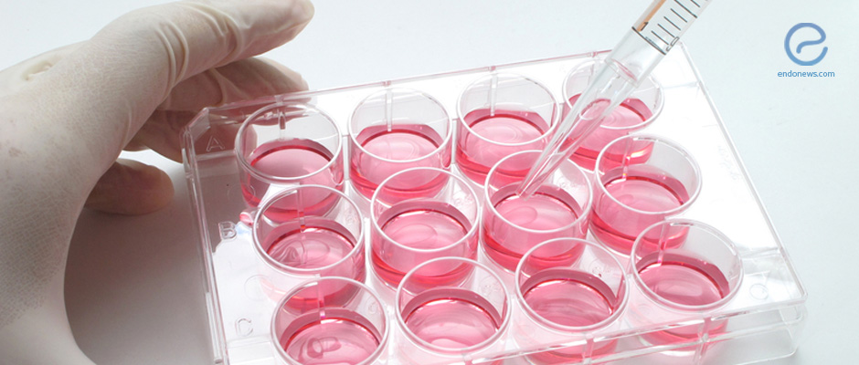
They did this by analyzing endometrial tissue samples obtained from women with and without endometriosis who underwent surgery and growing them in the laboratory under different conditions.
The samples analyzed a total of 80 women (40 with endometriosis and 40 without) between October 2015 and October 2016. The scientists grew the tissue samples in the laboratory in either a low or normal oxygen setting. They also exposed them to TGF-β1 and a molecule that blocks the activity of TGF-β1.
They then analyzed the levels of messenger RNA (mRNA) and protein of TGF-β1, VEGF, and hypoxia-inducible factor-1α (HIF-1α), a protein that is highly expressed in low oxygen. Messenger RNA is the molecular copy of a gene that will be made into a protein in the cell.
They found that the expression of all three molecules (TGF-β1, VEGF, and HIF-1α) were significantly higher, both at the mRNA and protein level, in tissues obtained from women with endometriosis compared to those in normal endometrial tissue.
When they grew the cells in a low oxygen environment, the expression of VEGF increased by 43.8 percent and when they treated them with TGF-β1, the expression of VEGF mRNA increased by 12.5 percent.
Importantly, when low oxygen levels were combined with TGF-β1 treatment, the expression of VEGF increased by a staggering 87.5 percent.
The treatment of the cells with the TGF-β1 inhibitor resulted in no difference in the expression of VEGF but low oxygen levels led to an increase of 41.2 percent increase in the expression of VGEF even in the presence of the inhibitor molecule.
Low oxygen levels increased the expression of HIF-1α, as well as that of TGF-β1 and TGF-β1 itself, increased the expression of HIF-1α.
The researchers concluded that TGF-β1 and hypoxia are having an additive effect in endometriosis at the transcriptional level.
Research Source: https://www.ncbi.nlm.nih.gov/pubmed/28397725
TGF-?1 hypoxia VGEF

