New Model Can Help Understand How Endometriosis-Associated Ovarian Cancer Develops
Jun 30, 2023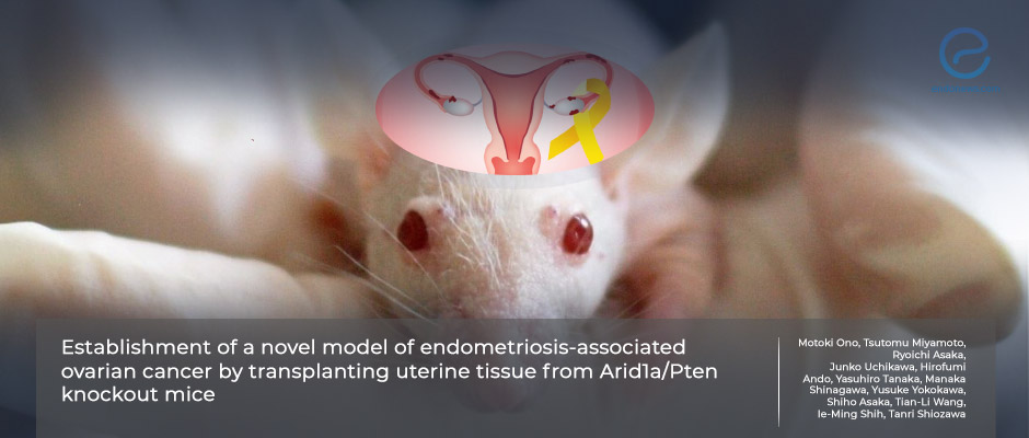
A new mouse model that can help understand the mechanisms underlimning the development of endometriosis-associated ovarian cancer was developed.
Key Points
Highlights:
- A new mouse model of endometriosis-associated ovarian cancer was recently developed.
Importance:
- The model can be useful in understanding the mechanisms of endometriosis-associated ovarian cancer development.
What’s done here:
- The team transplanted pieces of uterus from donor mice onto the ovarian surface or peritoneum of recipient conditionally knock-out mice, and then knocked out ARID1A and/or PTEN genes in turn.
Key results:
- When ARID1A only was knocked out, there were no histological changes in the endometriotic cysts of the recipient mice.
- When PTEN only was knocked out, the epithelial lining of the endometriotic cysts acquired a stratified architecture and nuclear atypia, which may correspond to atypical endometriosis.
- When both ARID1A and PTEN were knocked out papillary and cribriform structures and nuclear atypia were observed in about half of the peritoneal and ovarian endometriotic cysts similar to endometriosis-associated ovarian cancer.
Lay Summary
Researchers from Japan and the US developed a new mouse model in collaboration, that can be useful to better understand how endometriosis-associated ovarian cancer develops. This new model can help to assess new treatment options for both diseases.
The team led by Dr. Tanri Shiozawa from the Department of Obstetrics and Gynecology, Shinshu University School of Medicine, in Matsumoto, Japan created the model along with the collaboration of Molecular Genetics Laboratory of Female Reproductive Cancer of Johns Hopkins University, by transplanting pieces of uterus from donor mice onto the ovarian surface or peritoneum of recipient conditional knock-out mice. They then knocked out ARID1A and/or PTEN in the endometrial cells of the donor animals and analyzed the results. This is because these proteins as well as PIK3CA have been shown to play a role in ovarian cancer associated with endometriosis.
When only ARID1A was missing, the researchers saw that there were no histological changes in the endometriotic cysts of the recipient mice. But when only PTEN was missing, the epithelial lining of endometriotic cysts acquired a stratified architecture and nuclear atypia. This may correspond to atypical endometriosis.
When both ARID1A and PTEN were missing there were papillary and cribriform structures as well as nuclear atypia in the lining of 42% of peritoneal endometriotic cysts and half of ovarian endometriotic cysts. These were histologically similar to endometriosis-associated ovarian cancer.
“These results indicate that this mouse model is useful for investigating the mechanisms underlying the development of [endometriosis-associated ovarian cancer] and the related microenvironment,” the researchers concluded.
Research Source: https://pubmed.ncbi.nlm.nih.gov/37221199/
ovarian cancer endometriosis-associated ovarian cancer mouse model

