Immune cells in peritoneal endometriosis
Jul 15, 2022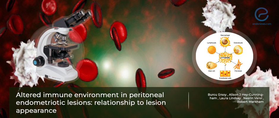
Immune cells may first be recruited to attack, but they might facilitate persistence and promote fibrosis.
Key Points
Highlights:
- How ectopic endometrial tissues attach to the peritoneum, persist, and develop has not been cleared yet.
- There is considerable variation in the appearances of endometriotic foci in relation to their ages, from early lesions to the red foci to progress to white scarred ones.
Importance:
- Different types of immune cells are increased in and around peritoneal endometriotic lesions in comparison to uninvolved peritoneum.
- Studying the lesion appearance with immune cells involved in each will widen our understanding of endometriosis pathogenesis.
What’s done here:
- A total of 30 tissue sample tissues from 28 patients undergoing laparoscopic excision of peritoneal endometriosis were recruited for this study.
- Immunohistochemical staining for mature and immature dendritic cells; effector, cytotoxic, regulatory T cells; B cells, and macrophages in endometriotic peritoneal lesions and surrounding tissue were assessed
- The assessment was performed on the histological sections of paraffin-embedded tissues.
Key results:
- Immune cells, especially in aggregates were more prominent in the earlier endometriosis lesions than the scarred ones.
- In tissues surrounding endometriotic foci, a greater number of immune cells were found in the youngest stage.
- Classifying immune cells in peritoneal endometriotic lesions may improve our understanding of the pathogenesis of persisting ectopic endometrial tissues.
Limitations of the study:
- The number of specimens included in this study could have been greater.
- The case number included in this study declined mainly due to exclusion of cases with combined color appearances (ie. red-black and black-white or even red-black-white).
- Other reasons for further exclusions from the study were as follows; surgically visualized lesions that could not be located within the paraffin tissue block microscopic sections or participants on hormonal treatment.
Lay Summary
Dr. Burcu Ersoy and associates from The University of Sydney, Australia, published their interesting study on surgically removed peritoneal endometriosis tissues in the journal named "F&S Science".
It is still a dilemma how ectopic endometrial tissues attach to the peritoneum, persist and develop. The endometriotic foci show a variation in color due to their age ranging from clear vesicles and red to black and finally scarred white ones. Previous studies did not consider lesion age when assessing the immune cell environment. The main aim of this study was to delineate for the first time, the immune environment of peritoneal endometriosis according to macroscopic appearances.
Different kinds of immune cells are increased in and around peritoneal endometriotic lesions in comparison to uninvolved peritoneum. Studying the lesion appearance with immune cells involved in each may widen our understanding of endometriosis pathogenesis.
Thirty lesion sample tissues from 28 patients undergoing laparoscopic excision of endometriosis were recruited for this prospective study from gynecological operations at Royal Prince Alfred Hospital, Sydney. The mean age of the patients was 32.6 years old. Immunohistochemical staining for mature and immature dendritic cells, different types of T cells, including effector, cytotoxic and regulatory ones; B cells, and macrophages in endometriotic peritoneal lesions and surrounding tissue were assessed on histopathological tissue sections.
Immune cell populations were present in and around peritoneal endometriotic lesions, with variations in numbers and distribution. Immature and mature dendritic cells, T cells, B cells, and macrophages were present in and around peritoneal endometriotic lesions. There were immune cell aggregates in most samples and the immune aggregates were 100-500 µm from the border of the endometriotic stroma and ranged in size from 100 µm in diameter, circular shapes up to 100-200 µm by 400 µm ovoid shapes. Immune cells, especially in aggregates were more often seen in the earlier lesions than the scarred ones. In surrounding tissues of endometriotic foci, a more crowded number of immune cells were found in the youngest stage.
These findings support the hypothesis that although immune cells may first be recruited to attack the endometriotic lesions, they might facilitate persistence and specifically promote fibrosis. Studies on functional outcomes are in need to elucidate the precise roles of immune cells in and around endometriosis for potential implications on possible treatment approaches.
Research Source: https://pubmed.ncbi.nlm.nih.gov/35559754/
peritoneal endometriosis immune cells immunohistochemistry

