Advanced Transvaginal Ultrasound for Endometriosis Mapping
Feb 13, 2026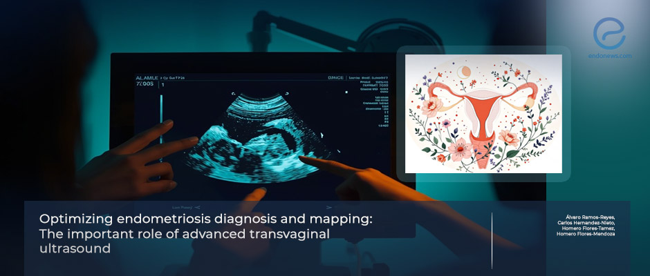
Preoperative Endometriosis Mapping Revisited: Advanced Ultrasound Demonstrates MRI-Level Precision
Key Points
Headline:
- Advanced transvaginal ultrasound (ATVUS) using the IDEA (International Deep Endometriosis Analysis) consensus enables systematic compartment-based mapping of endometriosis.
- ATVUS demonstrated strong concordance with MRI findings across pelvic compartments and was confirmed by surgical and histopathological correlation.
- Dynamic sonographic assessment, including sliding sign and mobility evaluation, provides additional clinically valuable information not obtainable through MRI.
Importance:
- Endometriosis diagnosis is frequently delayed by nearly 10 years, emphasizing the need for accessible and reliable first-line imaging tools.
- Accurate preoperative mapping improves surgical planning, reduces incomplete excision, and supports multidisciplinary surgical approaches.
- MRI remains highly accurate but may be limited by cost and accessibility, particularly in resource-constrained healthcare systems.
What's Done here?
- This 3 case-series report presents a case series of three patients evaluated with both ATVUS and MRI for endometriosis diagnosis and mapping.
- ATVUS was performed using a standardized IDEA consensus-based protocol assessing middle, posterior, and anterior pelvic compartments.
- Imaging findings were directly correlated with surgical observations and histopathological confirmation.
Key Results:
- ATVUS successfully identified ovarian endometriomas, uterosacral ligament nodules, rectosigmoid lesions, and posterior cul-de-sac obliteration with accuracy comparable to MRI.
- MRI detected additional superficial peritoneal implants and subtle ureteral involvement in selected cases.
- Dynamic ultrasound markers such as ovarian immobility and sliding sign accurately predicted adhesion severity and compartment involvement.
Strengths and limitations:
- Strengths are: the standardized IDEA-based compartmental imaging approach enhances reproducibility and clinical applicability; and the direct surgical and histological validation strengthens diagnostic reliability.
- Limitatioms are: very small sample size limits generalizability; highly operator-dependency of ATVUS, requiring specialized expertise.
From the Editor-in-Chief – EndoNews
"The growing recognition of endometriosis as a structurally complex and compartment-driven disease has shifted attention toward imaging strategies that move beyond lesion detection alone and toward functional and anatomical mapping. This study reinforces the clinical relevance of standardized, compartment-based imaging using advanced transvaginal ultrasound, particularly when performed under the framework of the International Deep Endometriosis Analysis (IDEA) consensus.
The findings support an important evolving paradigm: endometriosis should be understood not only as a distribution of lesions but as a disease interacting with anatomical planes, connective tissue scaffolding, and neurovascular networks. Lesions located within or adjacent to supportive ligaments, the rectovaginal septum, or deep posterior compartment structures often coincide with regions of dense neural distribution and fibrotic remodeling, which are increasingly recognized as key contributors to pain generation and disease persistence. Preoperative identification of disease within these functionally critical compartments therefore extends beyond surgical logistics and may directly influence long-term patient outcomes.
Advanced transvaginal ultrasound offers unique advantages in this context. Unlike static imaging modalities, its dynamic evaluation allows real-time assessment of tissue mobility, adhesion patterns, and organ sliding. These functional markers may serve as indirect indicators of fibrosis and neural involvement, providing clinically meaningful insights that complement structural lesion detection. The study further demonstrates that, when systematically applied, ATVUS can achieve diagnostic performance comparable to MRI across multiple pelvic compartments while maintaining superior accessibility and cost-effectiveness.
However, the conclusions should be interpreted within the context of the study design. The analysis is based on a very small case series including only three patients, which limits generalizability and prevents definitive comparisons between imaging modalities. Larger prospective and multicenter studies will be essential to confirm reproducibility and define the precise clinical role of ATVUS within diagnostic algorithms.
The results also highlight an essential limitation that remains central to ultrasound-based endometriosis imaging: operator dependency. The diagnostic strength of ATVUS is intrinsically linked to specialized training, structured protocols, and continuous surgical correlation. Expanding formalized education in IDEA-based imaging will therefore be critical for translating these benefits into broader clinical practice.
Importantly, this work contributes to the growing movement toward precision mapping in endometriosis. As surgical strategies increasingly emphasize complete excision while preserving function and minimizing complications, high-resolution preoperative compartment mapping may become indispensable. Future investigations integrating imaging findings with neuroanatomical, histopathological, and symptom-based data may further clarify how lesion location within specific pelvic compartments correlates with pain phenotypes and disease progression.
In this evolving landscape, advanced ultrasound should be viewed not as a replacement for MRI but as a complementary, highly adaptable imaging modality capable of bridging diagnostic accessibility with surgical precision. The continued refinement of standardized imaging frameworks such as the IDEA consensus will likely play a pivotal role in advancing personalized care for patients with endometriosis."
Lay Summary
Delayed diagnosis and incomplete disease mapping remain major challenges in the management of endometriosis, often contributing to prolonged symptoms and complex surgical outcomes. Improving preoperative identification of lesion location and disease extent is therefore critical for individualized treatment planning.
In a study published in the International Journal of Gynecology & Obstetrics, Dr. Álvaro Ramos-Reyes and colleagues evaluated Advanced transvaginal ultrasound (ATVUS) for endometriosis mapping, an advanced ultrasound technique performed using the International Deep Endometriosis Analysis (IDEA) consensus, an internationally standardized examination protocol that guides clinicians to systematically assess pelvic anatomical compartments.
By dividing the pelvis into structured regions—including the ovaries, bowel, bladder, and supporting ligaments—IDEA-based ATVUS for endometriosis mapping allows clinicians to generate a detailed map of disease distribution before surgery.
Compartment-based mapping is particularly important because endometriosis frequently develops near connective tissue planes and nerve-rich anatomical structures that are strongly associated with chronic pelvic pain and complex symptom patterns.
The researchers compared IDEA-guided ATVUS with magnetic resonance imaging in three patients and confirmed imaging findings through surgery and tissue examination.
IDEA-based ATVUS demonstrated diagnostic performance comparable to MRI across multiple pelvic compartments while also providing real-time evaluation of organ mobility and adhesions, features that cannot be directly assessed by MRI but are highly relevant for surgical planning.
Because ultrasound is widely available, cost-effective, and well tolerated, expanding training in standardized IDEA-based ATVUS for endometriosis mapping may help shorten diagnostic delays and improve access to specialized care.
Improved preoperative mapping may ultimately contribute to more complete surgical treatment and improved symptom control for individuals living with endometriosis.

